

Capture Every Detail with Moticam Pro S7 MONO Featuring Sony Pregius Sensor
Motic America
Read Article
Free Ground Shipping on Orders Over $300 Across USA and CANADA


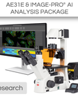
This Research Package includes:
*Advanced AI software is available for this package
The AE31E Trinocular is a live cell microscope platform for advanced microbiology. The powerful LWD CCIS© Phase objectives are designed for the inspection of native cell cultures. Lab technicians save time by benefitting from a clever illumination concept. The instrument can be upgraded with Fluorescence for research tasks, while the “Light memory” function and the Auto ON/OFF mode enables a fast and safe throughput of samples.
The Hi-Sensitives - Moticam ProS5 Plus is characterized by a large 2/3' sCMOS sensor and a Global Shutter for speedy handling of moving phenomena. This camera line takes Sensitivity as a priority: Fluorescence, Polarization, and Darkfield are the applications the engineers had in mind.
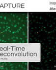
Unlock the full potential of your fluorescence microscope with the Image-Pro ® - Life Science Fluorescence Package for Motic, developed in partnership with Media Cybernetics. This all-in-one solution allows users to capture mixed sets of fluorescence and brightfield channels, then deconvolve them using AutoQuant Real-Time Deconvolution. Combining advanced Machine Learning technology with a comprehensive suite of protocols provides precise segmentation, classification, and automated measurements from 2D fluorescence images.
This package also includes a 12-month subscription to a Standard Success Plan from Media Cybernetics, providing ongoing technical assistance and software onboarding so you can focus on the work that truly matters.
| Model | AE31E Trinocular |
| Observation tube | Trinocular head, Siedentopf type, 360° swiveling |
| Optical system | Colour Corrected Infinity Optical System (CCIS®) |
| Condenser | Focusable and centerable ELWD condenser N.A. 0.30 (WD 72mm) |
| Diaphragm | Iris diaphragm |
| Diopter adjustment | On both eyepieces, +/- 5 diopter |
| Eyepieces | Widefield WF10X/22mm with diopter adjustment |
| Fine focus precision | 2µm |
| Fluorescence | Optional HBO |
| Focus mechanism | Coaxial coarse and fine focusing system with tension adjustment |
| Focusing stroke | 10mm |
| Free working distance | Working distance without condenser 231mm |
| Illumination features | Auto-OFF and Light Memory function |
| Illumination interchangeability | Quartz halogen 6V/30W or LED 3W |
| Inclination | 45° inclined |
| Interpupillary distance | 48-75mm |
| Nosepiece | Left side facing quintuple |
| Objective classification | CCIS® Plan Achromatic, DIN |
| Objective mounting thread | W 4/5"x1/36" (RMS standard) |
| Objectives | 4X/0.10 (WD 12.6mm), PH10X/0.25 (WD 4.1mm), LWD PH20X/0.30 (WD 4.7mm), [Optional] LWD PH40X/0.50 (WD 3.0mm) |
| Phase contrast | Slider |
| Stage | Plain stage with metal and glass stage inserts |
| Stage size | 200x239mm |
| Stand type | Inverted |
| Standard contrast technique | Brightfield, phase contrast |
| Transmitted illumination | Koehler Quartz halogen 6V/30W with intensity control |
| Transformer | Internal |
| Power supply | 100-240V (CE) |
| Accessories included | Dust cover, power cord, Allen key, blue, green interference and ground glass filters, phase ring Ph1, centering telescope, centering keys, spare fuse |
| Dimensions LxWxH | 556x200x529mm |
| Weight Net | 11.7kg |
| Model | Moticam ProS5 Plus |
| Sensor type | sCMOS |
| Sensor size | 2/3" |
| Imaging area | 11.1mm (Diagonal) |
| Capture resolution info | 5MP |
| Live display mode through USB (pixels) | 2448x2048, 1224x1024 |
| Pixel size | 3.45x3.45μm |
| Scan mode | Progressive |
| Shutter mode | Global Shutter |
| Data transfer | USB 3.1 |
| Max. frames per second (fps*) | 2448x2048 @ 68.3fps, 1224x1024 @ 175.8fps |
| Exposure time | 7μsec to 2 sec |
| Operating temperature | From -10 to +60 Degrees Celsius non condensing |
| Sensitivity | 1146mV(G) @ 1/30 sec |
| Support device | TWAIN, SDK and DirectShow Driver |
| Supported OS | Microsoft Windows 7/8/10, MAC OSX, Linux or higher |
| Minimum computer requirements | 2GHz dualcore - RAM memory 2GB - Video memory min. 512 MB |
| Lens mount | C-Mount |
| Software | Motic Images Plus 3.0 for Windows, OSX and Linux |
| Functions | Still image and video capture, live and still image measurement, image adjustments, white balance, automatic and manual exposure, individual objective calibration system |
| Power supply | 5V (from USB Port) |
| Package includes | CS ring adaptor, USB 3.1 cable, Motic 4-dot calibration slide, Motic Images Plus 3.0 for Windows/OSX/Linux |
| (*) Frames per second under optimal lighting conditions and in compliance with computer technical requirements. | |
|
|
Image-Pro® Software Package by Media Cybernetics
|
| Software Key Features | Standard Package | AI-Powered Package |
|
AI Deep Learning-Powered Analysis For superior 2D image segmentation accuracy over Machine Learning and other methods, this library of Pre-Trained AI models delivers instant segmentation results for a wide range of applications.
|
✓ | |
|
AI Deep Learning Model Training Fine tune the existing Pre-Trained models or build your own for custom segmentation of the most challenging images.
|
✓ | |
|
Machine Learning-Powered Analysis Leverage sophisticated pixel classification algorithms for accurate segmentation and classification, ensuring automated and reliable measurements from 2D images.
|
✓ | ✓ |
|
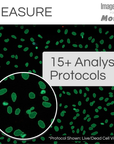
Analysis Protocols for guided analysis Streamline your research with a comprehensive suite of protocols designed for efficient, repeatable analysis across various life science applications—all within a simple, step-by-step workflow.
Includes: Angiogenesis, Cell Apoptosis, Autophagy, Confluence, Cell Count, Cell Morphology, Cell Proliferation, Transfection, Colocalization, Lipid Droplets, Live/Dead, Neurite Outgrowth, Nuclei, Ring Regions, Translocation, and Wound Healing. |
✓ | ✓ |
|
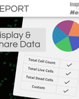
Data Visualization and Reporting Choose from 144+ measurements to display in tables and graphs, generate custom reports, and export data to Excel, CSV, or PDF. Easily share individual measurements and statistics with collaborators.
|
✓ | ✓ |
|
Image Processing Correct background illumination, tile and stitch large areas, colorize your images, enhance with filters, align and register images, and more. Choose from a helpful suite of options to prepare your images for analysis or publication.
|
✓ | ✓ |
|
Multi-Channel Fluorescence Capture Control the LUMOS LED light source with easy guided prompts to capture multiple fluorescence channels into a single multi-channel image, with Auto Exposure, and Real-Time Deconvolution.
|
✓ | ✓ |
|
Image Preview and Capture Designed to control the Moticam camera, you can now automatically preview a live image and effortlessly capture LIVE Tiling, LIVE EDF, LIVE HDR, Timelapse movies, and beautiful still images.
|
✓ | ✓ |
Check compatibility with Image-Pro
| Different packages have varying system requirements. Check your computer’s specifications against the system requirements for Image-Pro software. | See System Requirements | |
Microscope: AE31E Trinocular
Camera: Moticam ProS5 Plus
Install app