

Capture Every Detail with Moticam Pro S7 MONO Featuring Sony Pregius Sensor
Motic America
Read Article
Free Ground Shipping on Orders Over $300 Across USA and CANADA


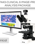

This Clinical Package includes:
*Advanced AI software is available for this package
The PA43BIO (Pathology) Trinocular is a perfect workhorse for pathologist. This microscope has a simple design and is very ease to use, but also does not compromise on quality and ergonomic features. It is a microscope meant to be used for long periods of time, with the highest quality of experience. The modularity of the PA43 microscopes offers you flexibility in choosing a configuration that suits your usage. It has a strong selection of contrast accessories that are available and easily upgradable to meet different requirements.
The Hi-Res Partner - Moticam S6 is designed for those application fields which need higher resolution. Presentations to a greater audience through video projector and reports in hardcopy are performed with ease. With 6 million pixels and a fast data readout, the performance is remarkable!
Transform your life sciences research with the Image-Pro® - Life Science Package for Motic, developed in partnership with Media Cybernetics. This versatile solution combines advanced Machine Learning technology with a comprehensive suite of protocols to provide precise segmentation, classification, and automated measurements from 2D images. Perfect for a wide range of life science applications, this package enhances your analytical capabilities with efficiency and accuracy.
This package also includes a 12-month subscription to a Standard Success Plan from Media Cybernetics, providing ongoing technical assistance and software onboarding so you can focus on the work that truly matters.
| Model | PA43BIO (Pathology) |
| Optical System | Scalable Infinity-Corrected Optical System (CCIS®) |
| Observation tube | Tilting Trinocular Inverted Image Tube Head |
| Inclination | 15-45° inclined |
| Trinocular light split | 100:0/50:50/0:100 |
| Interpupillary distance | 55-75mm |
| Eyepiece | Widefield WF10X/22mm with diopter adjustment |
| Diopter adjustment | On both eyepieces, +/- 5 diopter |
| Nosepieces | Coded Quintuple (5) revolving nosepiece |
| Objectives |
• Plan UC 2X/0.05, W.D=7.2mm, CG=0.17 • Plan UC 4X/0.1, W.D=30.5mm,CG=0.17 • Plan UC 10X/0.25, W.D=17.4mm,CG=0.17 • Plan UC 20X/0.45,W.D=0.8mm,CG=0.17 • Plan UC 40X/0.65,W.D=0.6mm,CG=0.17 |
| Condenser | Achromat Swing-out Condenser, N.A.0.90 / 0.13 |
| Stage | Rackless stage |
| Stage size | 180x170mm |
| Travel range X&Y | 80x55mm |
| Illumination | • LED illuminator • Halogen lamp drawer |
| Transmitted illumination | Koehler Illumination ,Integrated power supply Halogen 6V/30W and LED 3W |
| Model | Moticam S6 |
| Sensor type | sCMOS |
| Sensor size | 1/1.8" |
| Imaging area | 8.92mm (Diagonal) |
| Capture resolution info | 6MP |
| Live display mode through USB (pixels) | 3072x2048, 1536x1024 |
| Pixel size | 2.4x2.4μm |
| Scan mode | Progressive |
| Shutter mode | Rolling Shutter |
| Data transfer | USB 3.1 |
| Max. frames per second (fps*) | 3072x2048 @ 30fps, 1536x1024 @ 50fps |
| Exposure time | 16 μsec to 2 sec |
| Operating temperature | From -10 to +60 Degrees Celsius non condensing |
| Sensitivity | 425mV(G) @ 1/30 sec |
| Support device | TWAIN, SDK and DirectShow Driver |
| Supported OS | Microsoft Windows 7/8/10, MAC OSX, Linux or higher |
| Minimum computer requirements | 2GHz dualcore - RAM memory 2GB - Video memory min. 512 MB |
| Lens mount | C-Mount |
| Software | Motic Images Plus 3.0 for Windows, OSX and Linux |
| Functions | Still image and video capture, live and still image measurement, image adjustments, white balance, automatic and manual exposure, individual objective calibration system |
| Power supply | 5V (from USB Port) |
| Package includes | CS ring adaptor, USB 3.1 cable, Motic 4-dot calibration slide, Motic Images Plus 3.0 for Windows/OSX/Linux |
| (*) Frames per second under optimal lighting conditions and in compliance with computer technical requirements. | |
|
|
Image-Pro® Software Package by Media Cybernetics
|
| Software Key Features | Standard Package | AI-Powered Package |
|
AI Deep Learning-Powered Analysis For superior 2D image segmentation accuracy over Machine Learning and other methods, this library of Pre-Trained AI models delivers instant segmentation results for a wide range of applications.
|
✓ | |
|
AI Deep Learning Model Training Fine tune the existing Pre-Trained models or build your own for custom segmentation of the most challenging images.
|
✓ | |
|
Machine Learning-Powered Analysis Leverage sophisticated pixel classification algorithms for accurate segmentation and classification, ensuring automated and reliable measurements from 2D images.
|
✓ | ✓ |
|
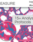
Analysis Protocols for guided analysis Streamline your research with a comprehensive suite of protocols designed for efficient, repeatable analysis across various life science applications—all within a simple, step-by-step workflow.
Includes: Angiogenesis, Cell Apoptosis, Autophagy, Confluence, Cell Count, Cell Morphology, Cell Proliferation, Transfection, Colocalization, Lipid Droplets, Live/Dead, Neurite Outgrowth, Nuclei, Ring Regions, Translocation, and Wound Healing. |
✓ | ✓ |
|
Data Visualization and Reporting Choose from 144+ measurements to display in tables and graphs, generate custom reports, and export data to Excel, CSV, or PDF. Easily share individual measurements and statistics with collaborators.
|
✓ | ✓ |
|
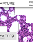
Image Processing Correct background illumination, tile and stitch large areas, colorize your images, enhance with filters, align and register images, and more. Choose from a helpful suite of options to prepare your images for analysis or publication.
|
✓ | ✓ |
|
Image Preview and Capture Designed to control the Moticam camera, you can now automatically preview a live image and effortlessly capture LIVE Tiling, LIVE EDF, LIVE HDR, Timelapse movies, and beautiful still images.
|
✓ | ✓ |
Check compatibility with Image-Pro
| Different packages have varying system requirements. Check your computer’s specifications against the system requirements for Image-Pro software. | See System Requirements | |
Microscope: PA43BIO (Pathology)
Camera: Moticam S6
Install app