

Capture Every Detail with Moticam Pro S7 MONO Featuring Sony Pregius Sensor
Motic America
Read Article
Free Ground Shipping on Orders Over $300 Across USA and CANADA


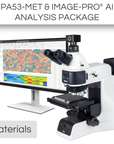
This Material Science Package includes:
*Advanced AI software is available for this package
The PA53MET-BD is superior solution for industrial quality control and analysis of opaque materials. Crisp and clear images are guaranteed thanks to the newly designed Semi-Apochromatic Brightfield and Darkfield objectives. The eyepieces have a 25mm FOV, ahead of any standard. The powerful 100W Halogen illumination features the ECO Function, a power-saving mode. The quintuple coded nosepiece recalls the last light intensity used for a smoother workflow. The incident illumination provides a variety of Contrast Methods: Brightfield and Darkfield are the standards, DIC is a powerful optional tool for the visualization of scratches or other tiny surface defects of opaque samples. The PA53MET BD is a pure incident illumination model for reflective materials.
The Hi-Res Partner - Moticam S6 is designed for those application fields which need higher resolution. Presentations to a greater audience through video projector and reports in hardcopy are performed with ease. With 6 million pixels and a fast data readout, the performance is remarkable!
Enhance your materials research with the Image-Pro ® - Materials Package for Motic, developed in partnership with Media Cybernetics. This comprehensive solution integrates advanced Machine Learning technology with a detailed set of protocols to provide precise segmentation, classification, and automated measurements from 2D images. Designed for diverse materials applications, this package boosts your analytical capabilities with efficiency and accuracy.
| Model | PA53MET-BD |
| Observation tube | Trinocular head, Siedentopf type |
| Optical system | Color Corrected Infinity Optical System (CCIS®) |
| Inclination |
20° inclined |
| Trinocular light split | 100:0/0:100 erect image |
| Interpupillary distance | 50-75mm |
| Diopter adjustment | On both eyepieces, +/- 4 diopter |
| Eyepieces | Widefield WF10X/25mm with diopter adjustment |
| Intermediate body | Epi-illuminator quartz halogen 100W with integrated field and aperture diaphragms, slots for filters and filters ND6, LBD |
| Nosepiece | Reversed quintuple, coded for brightfield and darkfield objectives with DIC slot |
| Objective classification | CCIS® Plan S-Apochromatic BD |
| Objectives | • 5X/0.15 (WD 20mm) • 10X/0.3 (WD 12mm) • 20X/0.45 (WD 3mm) • 50X/0.8 (WD 1mm) |
| Objective mounting thread | M26X0.706 |
| Stand type | Upright |
| Stage | Mechanical stage, hard coated with built-in low position coaxial stage control |
| Stage size | 210x170mm |
| Travel range X&Y | 104x102mm (4"x4") |
| Focus mechanism | Coaxial coarse and fine focusing system with tension adjustment |
| Fine focus precision | 1µm |
| Focusing stroke | 29.5mm - Coarse: 17.7mm/revolution - Fine: 0.1mm revolution (1µm scale) |
| Upper limit stop | Upper limit stop preset but adjustable |
| Incident illumination | Köhler Quartz halogen 12V/100W for BF, DF, DIC, POL with intensity control |
| Illumination features | Power saving mode ECO function, LED voltage indicator and Intelligent Light function |
| Transformer | Internal |
| Power supply | 110-240V (CE) |
| Dimensions LxWxH | 572x246x514mm |
| Weight Net | 19kg |
| Contrast techniques | |
| Standard contrast technique | Brightfield |
| Polarization | Optional slider reflected |
| Darkfield | Integrated |
| Modulation contrast | Optional slider reflected |
| Model | Moticam S6 |
| Sensor type | sCMOS |
| Sensor size | 1/1.8" |
| Imaging area | 8.92mm (Diagonal) |
| Capture resolution info | 6MP |
| Live display mode through USB (pixels) | 3072x2048, 1536x1024 |
| Pixel size | 2.4x2.4μm |
| Scan mode | Progressive |
| Shutter mode | Rolling Shutter |
| Data transfer | USB 3.1 |
| Max. frames per second (fps*) | 3072x2048 @ 30fps, 1536x1024 @ 50fps |
| Exposure time | 16 μsec to 2 sec |
| Operating temperature | From -10 to +60 Degrees Celsius non condensing |
| Sensitivity | 425mV(G) @ 1/30 sec |
| Support device | TWAIN, SDK and DirectShow Driver |
| Supported OS | Microsoft Windows 7/8/10, MAC OSX, Linux or higher |
| Minimum computer requirements | 2GHz dualcore - RAM memory 2GB - Video memory min. 512 MB |
| Lens mount | C-Mount |
| Software | Motic Images Plus 3.0 for Windows, OSX and Linux |
| Functions | Still image and video capture, live and still image measurement, image adjustments, white balance, automatic and manual exposure, individual objective calibration system |
| Power supply | 5V (from USB Port) |
| Package includes | CS ring adaptor, USB 3.1 cable, Motic 4-dot calibration slide, Motic Images Plus 3.0 for Windows/OSX/Linux |
| (*) Frames per second under optimal lighting conditions and in compliance with computer technical requirements. | |
|
|
Image-Pro® Software Package by Media Cybernetics
|
| Software Key Features | Standard Package | AI-Powered Package |
|
AI Deep Learning-Powered Analysis For superior 2D image segmentation accuracy over Machine Learning and other methods, this library of Pre-Trained AI models delivers instant segmentation results for a wide range of applications.
|
✓ | |
|
AI Deep Learning Model Training Fine tune the existing Pre-Trained models or build your own for custom segmentation of the most challenging images.
|
✓ | |
|
Machine Learning-Powered Analysis Leverage sophisticated pixel classification algorithms for accurate segmentation and classification, ensuring automated and reliable measurements from 2D images.
|
✓ | ✓ |
|
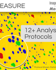
Analysis Protocols for guided analysis Streamline your research with a comprehensive suite of protocols designed for efficient, repeatable analysis across various materials applications—all within a simple, step-by-step workflow.
Includes: Composite Materials, Fiber Separation, Fiber Thickness, Grain, Layers, Particles Phase, Particle Size, Phases, and Pores. |
✓ | ✓ |
|
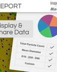
Data Visualization and Reporting Choose from 144+ measurements to display in tables and graphs, generate custom reports, and export data to Excel, CSV, or PDF. Easily share individual measurements and statistics with collaborators.
|
✓ | ✓ |
|
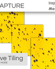
Image Processing Correct background illumination, tile and stitch large areas, colorize your images, enhance with filters, align and register images, and more. Choose from a helpful suite of options to prepare your images for analysis or publication.
|
✓ | ✓ |
|
Image Preview and Capture Designed to control the Moticam camera, you can now automatically preview a live image and effortlessly capture LIVE Tiling, LIVE EDF, LIVE HDR, Timelapse movies, and beautiful still images.
|
✓ | ✓ |
Check compatibility with Image-Pro
| Different packages have varying system requirements. Check your computer’s specifications against the system requirements for Image-Pro software. | See System Requirements | |
Microscope: PA53MET-BD
Camera: Moticam S6
Install app