

Capture Every Detail with Moticam Pro S7 MONO Featuring Sony Pregius Sensor
Motic America
Read Article
Free Ground Shipping on Orders Over $300 Across USA and CANADA


This Clinical Package includes:
*Advanced AI software is available for this package
The PA53BIO Trinocular is an upright laboratory microscope designed for research work in hospitals and life science laboratories. It features powerful Köhler Quartz Halogen 100W transmitted illumination and a 1µ fine focus precision. The IL Function, together with the quintuple coded nosepiece, memorizes the brightness level for each objective position.
The Hi-Res Partner - Moticam S6 is designed for those application fields which need higher resolution. Presentations to a greater audience through video projector and reports in hardcopy are performed with ease. With 6 million pixels and a fast data readout, the performance is remarkable!
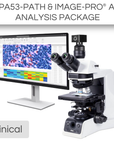
Transform your life sciences research with the Image-Pro® - Life Science Package for Motic, developed in partnership with Media Cybernetics. This versatile solution combines advanced Machine Learning technology with a comprehensive suite of protocols to provide precise segmentation, classification, and automated measurements from 2D images. Perfect for a wide range of life science applications, this package enhances your analytical capabilities with efficiency and accuracy.
This package also includes a 12-month subscription to a Standard Success Plan from Media Cybernetics, providing ongoing technical assistance and software onboarding so you can focus on the work that truly matters.
| Model | PA53BIO |
| Optical System | Colour Corrected Infinity Optical System (CCIS®) |
| Observation tube | Trinocular head, Siedentopf type |
| Inclination | 30° inclined |
| Trinocular light split | 100:0/20:80/0:100 |
| Interpupillary distance | 50-75mm |
| Diopter adjustment | On both eyepieces, +/- 4 diopter |
| Eyepieces | Widefield WF10X/23mm with diopter adjustment |
| Intermediate body | Simple intermediate |
| Nosepiece | Reversed quintuple, coded |
| Objective classification | CCIS® UC Plan Achromatic (Pb free) |
| Objectives | • 4X/0.10 (WD 30.5mm) • 10X/0.25 (WD 17.4mm) • 40X/0.65/S (WD 0.6mm) • 100X/1.25/S-Oil (WD 0.16mm) |
| Objective mounting thread | W 4/5"x1/36" (RMS standard) |
| Stand type | Upright |
| Stage | Mechanical stage with built-in low position rackless coaxial stage control and sample holder |
| Stage size | 220x170mm |
| Travel range X&Y | 80x55mm |
| Condenser | Focusable and centerable Abbe condenser N.A. 0.90/1.25 with slot for contrast sliders |
| Diaphragm | Iris diaphragm |
| Focus mechanism | Coaxial coarse and fine focusing system with tension adjustment |
| Fine focus precision | 1µm |
| Focusing stroke | 29.5mm - Coarse: 17.7mm/revolution - Fine: 0.1mm/revolution (1µm scale) |
| Upper limit stop | Upper limit stop preset but adjustable |
| Filter | ND6, ND25, LBD |
| Filter holder | Integrated filters |
| Transmitted illumination | Köhler Quartz halogen 12V/100W with intensity control |
| Illumination features | Power saving mode ECO function, LED voltage indicator and Intelligent Light function |
| Transformer | Internal |
| Power supply | 110-240V (CE) |
| Accessories included | Dust cover, power cord, Allen key, immersion oil (5ml) |
| Dimensions LxWxH | 576x247x457mm |
| Net weight | 19.2kg |
| Model | Moticam S6 |
| Sensor type | sCMOS |
| Sensor size | 1/1.8" |
| Imaging area | 8.92mm (Diagonal) |
| Capture resolution info | 6MP |
| Live display mode through USB (pixels) | 3072x2048, 1536x1024 |
| Pixel size | 2.4x2.4μm |
| Scan mode | Progressive |
| Shutter mode | Rolling Shutter |
| Data transfer | USB 3.1 |
| Max. frames per second (fps*) | 3072x2048 @ 30fps, 1536x1024 @ 50fps |
| Exposure time | 16 μsec to 2 sec |
| Operating temperature | From -10 to +60 Degrees Celsius non condensing |
| Sensitivity | 425mV(G) @ 1/30 sec |
| Support device | TWAIN, SDK and DirectShow Driver |
| Supported OS | Microsoft Windows 7/8/10, MAC OSX, Linux or higher |
| Minimum computer requirements | 2GHz dualcore - RAM memory 2GB - Video memory min. 512 MB |
| Lens mount | C-Mount |
| Software | Motic Images Plus 3.0 for Windows, OSX and Linux |
| Functions | Still image and video capture, live and still image measurement, image adjustments, white balance, automatic and manual exposure, individual objective calibration system |
| Power supply | 5V (from USB Port) |
| Package includes | CS ring adaptor, USB 3.1 cable, Motic 4-dot calibration slide, Motic Images Plus 3.0 for Windows/OSX/Linux |
| (*) Frames per second under optimal lighting conditions and in compliance with computer technical requirements. | |
|
|
Image-Pro® Software Package by Media Cybernetics
|
| Software Key Features | Standard Package | AI-Powered Package |
|
AI Deep Learning-Powered Analysis For superior 2D image segmentation accuracy over Machine Learning and other methods, this library of Pre-Trained AI models delivers instant segmentation results for a wide range of applications.
|
✓ | |
|
AI Deep Learning Model Training Fine tune the existing Pre-Trained models or build your own for custom segmentation of the most challenging images.
|
✓ | |
|
Machine Learning-Powered Analysis Leverage sophisticated pixel classification algorithms for accurate segmentation and classification, ensuring automated and reliable measurements from 2D images.
|
✓ | ✓ |
|
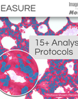
Analysis Protocols for guided analysis Streamline your research with a comprehensive suite of protocols designed for efficient, repeatable analysis across various life science applications—all within a simple, step-by-step workflow.
Includes: Angiogenesis, Cell Apoptosis, Autophagy, Confluence, Cell Count, Cell Morphology, Cell Proliferation, Transfection, Colocalization, Lipid Droplets, Live/Dead, Neurite Outgrowth, Nuclei, Ring Regions, Translocation, and Wound Healing. |
✓ | ✓ |
|
Data Visualization and Reporting Choose from 144+ measurements to display in tables and graphs, generate custom reports, and export data to Excel, CSV, or PDF. Easily share individual measurements and statistics with collaborators.
|
✓ | ✓ |
|
Image Processing Correct background illumination, tile and stitch large areas, colorize your images, enhance with filters, align and register images, and more. Choose from a helpful suite of options to prepare your images for analysis or publication.
|
✓ | ✓ |
|
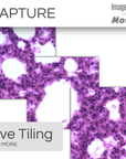
Image Preview and Capture Designed to control the Moticam camera, you can now automatically preview a live image and effortlessly capture LIVE Tiling, LIVE EDF, LIVE HDR, Timelapse movies, and beautiful still images.
|
✓ | ✓ |
Check compatibility with Image-Pro
| Different packages have varying system requirements. Check your computer’s specifications against the system requirements for Image-Pro software. | See System Requirements | |
Microscope: PA53BIO
Camera: Moticam S6
Install app