

Capture Every Detail with Moticam Pro S7 MONO Featuring Sony Pregius Sensor
Motic America
Read Article
Free Ground Shipping on Orders Over $300 Across USA and CANADA
Posted by Motic America on

Introduction
A coverslip also called a coverglass is usually made of thin pieces of borosilicate glass that are used to cover specimens viewed on a microscope slide. The thickness of the coverslip is critical for high magnification using “dry” objectives (no immersion oil) for optimum results in photomicrography. Coverslip are also made of quartz where enhanced UV (ultra violet) transparency is needed. New coverslips for confocal laser scanning microscopy for use with water immersion objectives have been developed called Cytop. These coverslips have a refractive index (1.35 to 1.4) close to water and are made of an acrylate resin (M Brenner et al. 2011). Plastic coverslips are primarily used in education and are not recommended for photomicrography or use with polarized light microscopy because they are birefringent. With fluorescence microscopy some plastic coverslips are auto-fluorescent. Plastic coverslips for general or educational use are lower in cost, unbreakable, and disposable. Coverslips come in different thickness; choosing the correct thickness of coverslip is one of the most important tasks in achieving high resolution and sharp photomicrographs. Using an incorrect thickness coverslip results in spherical aberrations which result in soft hazy images.
Most microscope objectives are designed for use with coverslips that are 0.17 mm thick when the specimen is in direct contact with the overlying coverglass. The 0.17 number is printed on the objective sleeve (see Fig. 1 above). Objectives of higher numerical aperture (NA) greater than 0.4 are more sensitive to the thickness of the coverslip than those of lower NA. Objectives starting at 20X NA 0.4 up to NA 0.95 (63X) are most sensitive to spherical aberration caused by incorrect coverslip thickness.
The thickness of 0.17 mm on the objective does not refer to just the coverslip thickness, but the combination of the coverslip thickness and the thickness of medium above the specimen (M. Kozbek 2001). Therefore thick specimens with large amounts of medium above the specimen require thinner coverslips. The mountant or aqueous solution can vary from 5 to 76 microns or more above the specimen (B. Neuhaus, T. Schmid and J. Riedel , 2017). Experienced microscopists generally choose No. 1.5 coverglasses that are 0.17 mm thick when the specimen is in contact with the coverslip or close to it. Some researchers and technicians grow cells directly on the coverslip and then mount them on a slide. I use No 1.5H coverglasses most of the time but I found that sometimes using thinner No. 1 coverslips are better due to varying amounts of overlying fluid. One way to reduce water or saline above a specimen is to draw the extra water out from below the coverslip by using a small piece of paper towel, filter paper, or allow time for evaporation (see below). This will result in sharper pictures most of the time.

Coverglasses are available in different thickness ranges: Number 0, 1, 1.5, 1.5H, 2, 3, and 4. Higher numbers indicate thicker coverglasses (see table below). The proper thickness for most objectives is 0.17mm (some older objectives were made for use with 0.18 mm coverglass). Some companies offer No 1.5H coverglasses with a tolerance of +\- 0.005 mm thick (H stands for high performance). These 1.5H coverslips cost about $100\10,000 but prices from different companies can vary by more than ten fold. Cover glasses are sold as round, square or rectangular in size depending on their intended use. Rectangular coverglasses are often used for blood smears or on hemocytometer slides, and I found their larger size is useful for scanning plankton. Round coverglasses are easier to “ring” i.e. seal them for long term storage and 22 x 22 mm square coverglasses are commonly used for other specimens.

J.G. Delly (1988, 2017) measured the thickness of coverglasses in a package and showed they have a normal distribution around the mean thickness specified. Some researchers measure the thickness of the coverglass with a digital micrometer for critical use. Measuring the coverglass thickness isn’t routinely done as it is time consuming, and the exact amount of fluid above the specimen is difficult to control accurately. However, researchers sometimes measure the coverslips thickness for confocal laser scanning microscopy or when the highest resolution and maximum quality images are required.
Table of Coverglass Thickness
| Coverslip # | Thickness (mm) |
| 0 | 0.085 – 0.13 |
| 1 | 0.13 – 0.16 |
| 1.5 | 0.16 – 0.19 |
| 1.5H | 0.170 +\- 0.005 |
| 2 | 0.19 – 0.23 |
| 3 | 0.24 – 0.35 |
| 4 | 0.43 – 0.65 |
Sometimes aquatic subjects I photograph with a microscope are not clear and sharp even when using 1.5H coverglasses and high NA objectives (40X and 63X), but as the water under the coverslip evaporates the coverslip comes into contact with the surface of the aquatic organisms and the image quality improves. I tested No 1 coverslips for live aquatic specimens and found that the images were often clear and crisp. Furthermore when I purchase prepared microscope slides e.g. plant specimens, many companies use No. 1 coverslips on their permanent slides. The reason is that the 0.17mm refers to both the combined specimen and the thickness of the mounting medium or saline above it. For this reason using the 40X and 63X dry objectives it is important to try to minimize the fluid above the specimen drawing excess water out from below the coverslip. When making permanent slides it is also important to minimize the amount of mounting fluid above the specimen.
With low power 2.5X, 4X and 10X objectives the thickness of the coverslip makes little difference in image quality because these objectives have a large depth of field. When using polarized light microscopy and crystals I achieve sharp crystal images with 10X or lower objectives with or without the use of a coverslip.


Is Coverslip Thickness important when using an Oil Immersion Lens?
No. 1 coverslips are frequently used to make permanent slides using a mounting medium like Canada Balsam with refractive index = 1.53 similar to that of glass. No. 1 coverslips are used so that the total thickness of the coverslip and mounting medium is close to 0.17 mm. One study found that putting a weight or using clothes pins to press the coverslip down during mounting with Canada Balsam resulted in thinner amounts of medium above the specimen and clearer images and recommends the use of No. 1 coverslips for permanent slides (G.W. Gill, 2013).
When using oil immersion objectives with permanent slides coverslip thickness is only of limited concern because the refractive index of the oil immersion fluid matches that of the coverglass and mountant. However, if a specimen is mounted in physiological saline or water where the refractive index is significantly lower, the thickness of the coverglass and overlying height of fluid is important so one should use No. 1.5 or No. 1 coverslips (T.J. Fellers, and M.W. Davidson). An alternative option is to mount specimens directly in oil immersion fluid if possible e.g. human hair - this can improve the resolution and clarity of the specimen.
For confocal laser scanning microscopy it is advisable to use high quality No. 1.5H coverslips. Inverted microscopes used for photomicrography require inverting the slides or using petri dishes with coverslip bottoms 0.17 mm thick for best results. Thicker coverslips No. 2-4 are sometimes used for observing whole mounts of arthropods and other invertebrates with lower magnification objectives and for counting yeast, blood cells and plankton.
Objectives with Correction Collars
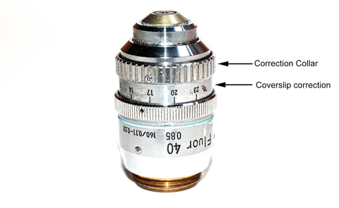
Objectives with correction collars can be used to correct for different thickness coverslips. These are usually high NA aperture objectives and expensive. One must be careful with these objectives as it is possible to break the coverslips with them while compensating. To use them you view your specimen via the eyepieces, fine focus on the subject and turn the objective collar until you achieve the sharpest image by trial and error. By turning the objective correction collar one can compensate for small differences in spherical aberration due to differences in coverslip thickness within a narrow range. I own one objective with a correction collar, a 40X Neofluor for fluorescence microscopy shown above.

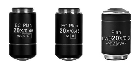
Some objectives are designed for use without a coverslip and they have a 0 on the objective sleeve, other objectives may have 1.1 mm and they are designed for water/agar medium. Some microscope companies offer multi-immersion objectives with a correction collar that can be used with immersion oil, glycerin and water. A few older microscopes have adjustable drawtubes so the mechanical tube length could be modified to accommodate different thickness coverglass. Sometimes pulling the microscope eyepieces out a bit on older microscopes changes the tube length and can produce sharper images when using the wrong thickness of coverglass.
For highest resolution you can choose to use oil immersion 40X and 63X objectives but the mountant must have the same refractive index of the glass coverslip to prevent spherical aberration. When the NA of the objective exceeds 1 you should also include oil immersion fluid between the condenser and microscope slide (J. G. Delly, 2017). The thickness of the glass microscope slide is also important but not as important as the coverslip thickness. Generally microscopes slides are between 1.0 to 1.2 mm thick with 1.0 mm being suitable for most subjects (J. G. Delly 1988).
Measuring Cover Glass Thickness
It is simple to measure coverslip thickness with a digital micrometer. I use a Neoteck digital micrometer with a resolution of 0.001 mm (cost $50 online). It is impractical to measure coverglass thickness routinely unless one is performing high resolution measurements or trying to detect single molecules. If your specimen is uniform in height, e.g. polystyrene beads used for determining resolution or calibration, then measuring the coverglass thickness so they are exactly 0.17 mm thick should provide optimum results. For most purposes selecting No.1.5 or No. 1 coverglasses will provide good results for general photomicrography provided the saline or water above the specimen is minimized.

Dirty coverslips
It is important that a microscope be kept clean of dust and dirt, including the camera sensor. Likewise it is essential that microscope slides and coverslips be clean. I examined coverslips from a variety of manufacturers and was surprised how dirty some of the coverslip surfaces were straight out of the box. I usually clean microscope slides using Windex™ and 95% isopropanol for routine work. I tried cleaning coverslips but found they often break during cleaning and it is tedious and time consuming. Some microscopes with darkfield, or epifluorescence microscopy require “super-clean” slides and coverglass. For cleaning you need a dust free location (fume or culture hood). Often slides and coverslips are immersed in IMHCl for about an hour, then rinsed several times with distilled water, followed by rinsing in 95% alcohol and dried. Slides are dried between clean Whatman paper or blown-dry and stored in a clean box. There are cleaning procedures available online from different labs. For examining cell cultures often the coverslips are coated with polylysine, fibronectin etc. and sterilized with UV or heat.
Summary and Conclusions
Factors influencing the sharpness and clarity of images viewed by a microscope are complex. Getting the maximum resolution with high dry microscope objectives (20X) NA 0.4 and above and with oil immersion objectives requires consideration of the coverglass thickness, the thickness of the medium above the specimen and the mediums refractive index. For general examination of aquatic microorganisms No. 1 and No 1.5 coverslips work well. No. 1 coverslips appear to be better for making permanent mounts of prepared tissues. For the sharpest images with high NA objectives of live specimens in water or saline minimize the amount of solution above the specimen by removing excess water from under the coverslip. No 1.5 H coverslips are high performance and recommended for photomicrography. Sometimes “little things” can make a big difference.
Related Products
| Models |  |
|
 |
| Features |
The BA310 is designed for the daily routine work in universities, clinics, laboratories, and life sciences or medical applications. |
The BA410E allows a profound diagnosis in pathology, hematology, and cytology with their requirements for best color fidelity. |
Panthera C2 is a new world-class level cased in a revolutionary technical future-orientated solution, now accessible for life sciences. |
Want to know which microscopes fit you best? Send us a message and our specialists are glad to help!
References
Some Sources of Cover glass
1) Zeiss No 1.5H, 2) Thor labs No 1.5 H, 3) Marienfield No 1.5H coverslips, 4) Thomas scientific plastic coverslips, 5) No 1.5 coverglass from China, 6) Muhawa cover glass from China precleaned 24 x 50 mm No 1 coverslips

Industrial and material sciences focus on studying the structure, properties, and performance of materials used in manufacturing, engineering, and product development. These fields involve close...

Microscopes are essential tools in biomedical research. These instruments help scientists, lab professionals, and researchers examine cells, tissues, bacteria, and other microscopic structures that are...