
Diatoms - Nature’s Jewels viewed with a Microscope
Motic America
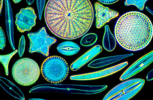
Diatoms are unicellular algae (Division Chrysophyta, Class Bacillariophyceae). Diatoms are microscopic in size, live in water, soil and moist en...
Read Article
Diatoms are unicellular algae (Division Chrysophyta, Class Bacillariophyceae). Diatoms are microscopic in size, live in water, soil and moist en...
Read Article


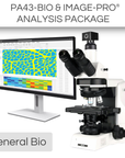


This General Bio Package includes:
*Advanced AI software is available for this package
The PA43BIO (Standard) Trinocular is a perfect workhorse for clinical routine work. This microscope has a simple design and is very ease to use, but also does not compromise on quality and ergonomic features. It is a microscope meant to be used for long periods of time, with the highest quality of experience. The modularity of the PA43 microscopes offers you flexibility in choosing a configuration that suits your usage. It has a strong selection of contrast accessories that are available and easily upgradable to meet different requirements.
The Hi-Res Partner - Moticam S6 is designed for those application fields which need higher resolution. Presentations to a greater audience through video projector and reports in hardcopy are performed with ease. With 6 million pixels and a fast data readout, the performance is remarkable!
Transform your life sciences research with the Image-Pro® - Life Science Package for Motic, developed in partnership with Media Cybernetics. This versatile solution combines advanced Machine Learning technology with a comprehensive suite of protocols to provide precise segmentation, classification, and automated measurements from 2D images. Perfect for a wide range of life science applications, this package enhances your analytical capabilities with efficiency and accuracy.
This package also includes a 12-month subscription to a Standard Success Plan from Media Cybernetics, providing ongoing technical assistance and software onboarding so you can focus on the work that truly matters.
| Model | PA43BIO (Standard) |
| Observation tube | Trinocular tube head |
| Eyepiece | N-WF10X/22mm |
| Revolving nosepiece | 5-position nosepiece |
| Objective | • Plan UC 4X/0.1, W.D=30.5mm • Plan UC 10X/0.25, W.D=17.4mm • Plan UC 40X/0.65, W.D=0.6mm • Plan UC 100X/1.25 Oil, W.D=0.16mm |
| Condenser | N.A 0.9/1.25 Abbe condenser with slider slot |
| Stage | Rackless stage |
| Transmitted illumination | Condensor (w/ field diaphragm) |
| Illumination | • LED module (high color temperature) • Halogen module |
| Filter | Blue filter |
| Model | Moticam S6 |
| Sensor type | sCMOS |
| Sensor size | 1/1.8" |
| Imaging area | 8.92mm (Diagonal) |
| Capture resolution info | 6MP |
| Live display mode through USB (pixels) | 3072x2048, 1536x1024 |
| Pixel size | 2.4x2.4μm |
| Scan mode | Progressive |
| Shutter mode | Rolling Shutter |
| Data transfer | USB 3.1 |
| Max. frames per second (fps*) | 3072x2048 @ 30fps, 1536x1024 @ 50fps |
| Exposure time | 16 μsec to 2 sec |
| Operating temperature | From -10 to +60 Degrees Celsius non condensing |
| Sensitivity | 425mV(G) @ 1/30 sec |
| Support device | TWAIN, SDK and DirectShow Driver |
| Supported OS | Microsoft Windows 7/8/10, MAC OSX, Linux or higher |
| Minimum computer requirements | 2GHz dualcore - RAM memory 2GB - Video memory min. 512 MB |
| Lens mount | C-Mount |
| Software | Motic Images Plus 3.0 for Windows, OSX and Linux |
| Functions | Still image and video capture, live and still image measurement, image adjustments, white balance, automatic and manual exposure, individual objective calibration system |
| Power supply | 5V (from USB Port) |
| Package includes | CS ring adaptor, USB 3.1 cable, Motic 4-dot calibration slide, Motic Images Plus 3.0 for Windows/OSX/Linux |
| (*) Frames per second under optimal lighting conditions and in compliance with computer technical requirements. | |
|
|
Image-Pro® Software Package by Media Cybernetics
|
| Software Key Features | Standard Package | AI-Powered Package |
|
AI Deep Learning-Powered Analysis For superior 2D image segmentation accuracy over Machine Learning and other methods, this library of Pre-Trained AI models delivers instant segmentation results for a wide range of applications.
|
✓ | |
|
AI Deep Learning Model Training Fine tune the existing Pre-Trained models or build your own for custom segmentation of the most challenging images.
|
✓ | |
|
Machine Learning-Powered Analysis Leverage sophisticated pixel classification algorithms for accurate segmentation and classification, ensuring automated and reliable measurements from 2D images.
|
✓ | ✓ |
|
Analysis Protocols for guided analysis Streamline your research with a comprehensive suite of protocols designed for efficient, repeatable analysis across various life science applications—all within a simple, step-by-step workflow.
Includes: Angiogenesis, Cell Apoptosis, Autophagy, Confluence, Cell Count, Cell Morphology, Cell Proliferation, Transfection, Colocalization, Lipid Droplets, Live/Dead, Neurite Outgrowth, Nuclei, Ring Regions, Translocation, and Wound Healing. |
✓ | ✓ |
|
Data Visualization and Reporting Choose from 144+ measurements to display in tables and graphs, generate custom reports, and export data to Excel, CSV, or PDF. Easily share individual measurements and statistics with collaborators.
|
✓ | ✓ |
|
Image Processing Correct background illumination, tile and stitch large areas, colorize your images, enhance with filters, align and register images, and more. Choose from a helpful suite of options to prepare your images for analysis or publication.
|
✓ | ✓ |
|
Image Preview and Capture Designed to control the Moticam camera, you can now automatically preview a live image and effortlessly capture LIVE Tiling, LIVE EDF, LIVE HDR, Timelapse movies, and beautiful still images.
|
✓ | ✓ |
Check compatibility with Image-Pro
| Different packages have varying system requirements. Check your computer’s specifications against the system requirements for Image-Pro software. | See System Requirements | |
Microscope: PA43BIO (Standard)
Camera: Moticam S6
Install app