
Diatoms - Nature’s Jewels viewed with a Microscope
Motic America
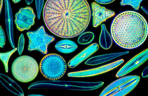
Diatoms are unicellular algae (Division Chrysophyta, Class Bacillariophyceae). Diatoms are microscopic in size, live in water, soil and moist en...
Read Article
Diatoms are unicellular algae (Division Chrysophyta, Class Bacillariophyceae). Diatoms are microscopic in size, live in water, soil and moist en...
Read Article

Posted by Motic America on
Motic Images Plus 3.1 Advanced Software is the most sophisticated imaging analysis software from Motic. It consists of two main programs: the capture interface which controls a Moticam camera and the captured image application program, and consists advanced features include image stitching and fluorescence image processing, which unlocking the potential of your microscope and enhancing your digital microscopy platform into the next level.

Motic Images Plus 3.1 Advanced Software
Motic Images Plus 3.1 Advanced Software includes all features and tools of the Motic Images Plus 3.0, with additional three different modules: Manual Segmentation, Motic Images Multi-focus and Motic Images Assembly.
| Manual Segmentation | The software allows segmentation of images manually with thresholds for red, green, blue and gray. It is also possible to perform single-point segmentation with up to eight different colour levels. Several filters such as Hi-Pass and Low-Pass as well as customization options are available. |
| Motic Images Multi-focus | This feature allows the user to capture images at different focal depths. The software will scan each layer and assemble a new image with all maximum value pixels thereby creating a single image where all layers are in focus. The program even automatically adjusts and compensates for any image shift when using Stereo Microscopy. |
| Motic Images Assembly | When looking at objects with high magnification, the field of view decreases. This feature will allow users to capture images at a high field of view as well as high magnification. Up to 100 images taken in one focal plane in a 10x10 format can be scanned by this software and assembled. All overlaps are recognized and individual images are automatically shifted into the right place. |
Other functions included are:
Want to know which microscopes fit you best? Fill in the form below and our specialists are glad to help!

Motic™ Instruments Inc., a global leader in microscope solutions, and Media Cybernetics Inc., an innovator in AI-powered image analysis software...

Motic is excited to announce the relocation of US office at Suite 197, 1500 Northlake Pass, Universal City, TX 78148, start operating on 12 December...